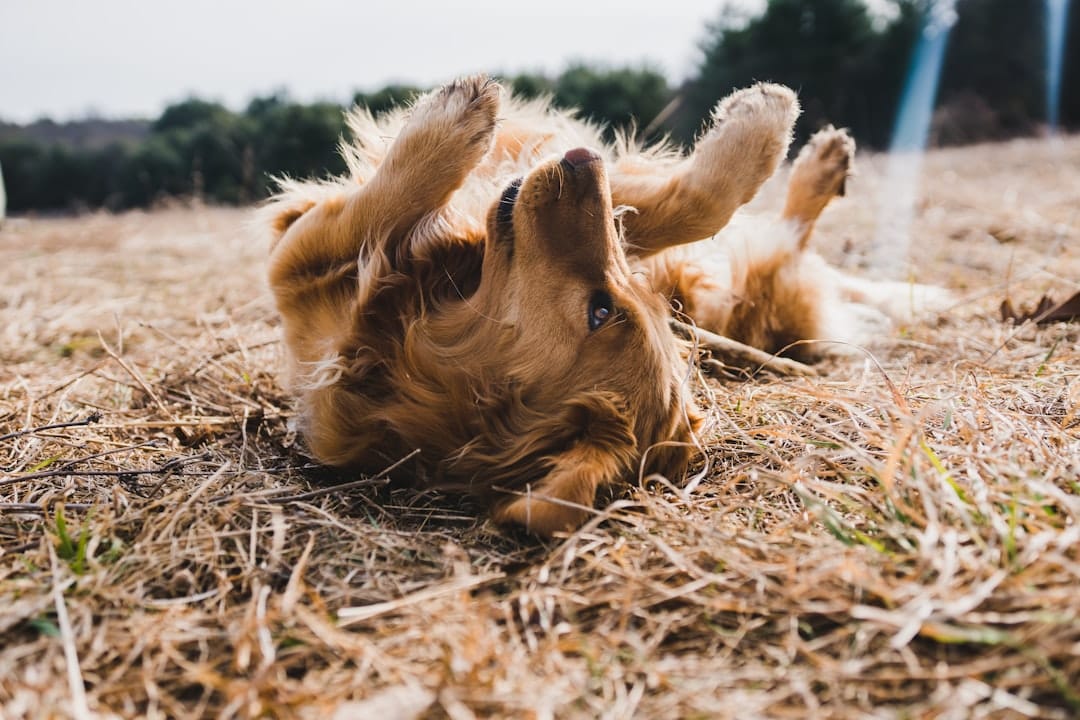
When Luna started leaving tufts of golden fur on the beige sofa last March, her owner Sarah initially dismissed it as seasonal shedding. But within weeks, the three-year-old Labrador developed angry pink patches on her belly and began obsessively licking her paws until the fur turned rust-colored. “I thought she just had dry skin,” Sarah recalls, “until I realized we were dealing with something that needed medical attention.”

If you’re searching for what do allergies look like in dogs, you’re likely noticing similar troubling changes in your own pup. Unlike humans who sneeze and get watery eyes, canine allergies often manifest through the skin and coat—making them harder to identify but impossible to ignore once they progress. This comprehensive 2026 guide will help you recognize the visual cues, behavioral shifts, and physical markers that distinguish allergic reactions from other common conditions.

The Visual Language of Canine Allergies
Understanding dog skin allergy treatment begins with recognizing how allergies physically present themselves. While symptoms vary based on the allergen type—environmental, food, or flea-related—most allergic dogs display a specific visual vocabulary that observant owners can learn to read.
Skin Manifestations: Beyond Basic Itching
The skin serves as the primary billboard for allergic distress in dogs. Unlike the isolated dry patches that come with winter weather, allergic skin reactions typically appear as symmetrical patterns of inflammation. You might notice erythema—medical terminology for redness—particularly in thinly furred areas like the groin, armpits, and inner thighs.
Chronic scratching creates secondary visual markers. Look for excoriations, which are linear scratch marks that break the skin surface, often accompanied by crusting or scabbing. Dogs with chronic allergies frequently develop lichenification, where the skin thickens and develops exaggerated texture resembling elephant skin, typically on the abdomen or flanks.
Hot spots (acute moist dermatitis) represent another visual red flag. These appear suddenly as circular, reddened, moist lesions that may ooze serum. While any dog can develop a hot spot from a single irritant, allergic dogs experience recurring hotspots in the same locations—often the cheeks, neck, or base of the tail.
Ear Signals: The Canary in the Coal Mine
Otitis externa—inflammation of the external ear canal—serves as one of the earliest visual indicators of allergies in many dogs. Examine your dog’s ears for a deepening pink or red hue inside the pinnae (ear flaps). Allergic ears often produce excessive brown, yellow, or black discharge that differs from healthy ear wax.
Chronic ear infections in allergic dogs create distinctive physical changes. The ear canals may narrow due to swelling, and the leather of the ear flaps can thicken. Some dogs develop a characteristic “ear hematoma”—a fluid-filled swelling on the ear flap caused by violent head-shaking and scratching.
Gastrointestinal Red Flags
While less visible externally, canine food allergy symptoms manifest through specific physical markers. Dogs with food allergies often develop perioral dermatitis—red, inflamed skin around the mouth and muzzle. You might notice frequent lip-licking, face-rubbing against furniture, or pawing at the muzzle after eating.
Chronic loose stools or mucus-coated feces indicate gastrointestinal involvement. Some allergic dogs develop “bilious vomiting syndrome” appearances—yellow foamy vomit—particularly in the morning. Weight loss may become visible along the spine and ribs despite normal food intake, indicating malabsorption issues.
Respiratory and Ocular Signs
Though less common than skin symptoms, respiratory allergies do occur in dogs. Look for bilateral clear nasal discharge (from both nostrils), reverse sneezing episodes that sound like honking, and conjunctival hyperemia—redness of the eye whites. Allergic dogs may develop tear staining—rust-colored tracks running from the inner corners of the eyes down the muzzle.
Flea Allergies vs. Environmental Allergies: Visual Comparison
Distinguishing between flea allergy dermatitis (FAD) and atopic dermatitis helps target treatment effectively. While both cause intense itching, their visual patterns differ significantly.
| Visual Marker | Flea Allergy Dermatitis | Environmental Allergies |
|---|---|---|
| Primary Location | Base of tail, inner thighs, lower back | Paws, face, ears, belly |
| Hair Loss Pattern | Symmetric bald patches, “rat tail” appearance | Patchy thinning, broken hairs from chewing |
| Skin Changes | Papules (small red bumps), flea dirt visible | Erythema, lichenification, hyperpigmentation |
| Secondary Signs | Scabs along dorsal midline | Salivary staining on paws (red-brown discoloration) |
| Seasonality | Year-round if fleas present, worse in warm months | Often seasonal (spring/fall) or year-round |
The Progression Timeline: Acute vs. Chronic Allergic Reactions
Recognizing best dog allergy relief strategies requires understanding whether you’re viewing acute or chronic manifestations.
Acute Reactions (Hours to Days): Hives (urticaria) appear as raised, circular welts resembling mosquito bites across the body. Facial swelling (angioedema) may cause the muzzle to look puffy or the eyelids to swell shut. These require immediate veterinary attention.
Subacute Phase (Days to Weeks): The skin transitions from red to purple-gray as pigment changes occur. Secondary yeast infections create a greasy feel and corn-chip odor, particularly in skin folds. The coat loses luster and may feel brittle.
Chronic Changes (Months to Years): Long-standing allergies remodel the skin permanently. Hyperpigmentation turns light-colored skin dark brown or black. The skin barrier becomes compromised, leading to frequent bacterial infections requiring antibiotic therapy. Some dogs develop calcinosis cutis—white, chalky calcium deposits in the skin—from prolonged steroid use.
When It’s Not Allergies: Visual Mimics to Rule Out
Several conditions resemble allergies but require different treatments. Mange (demodectic or sarcoptic) causes similar hair loss but typically features more scaling and less inflammation. Fungal infections like ringworm create circular lesions with central clearing, unlike the diffuse redness of allergies.
Hormonal imbalances such as hypothyroidism cause symmetric hair loss on the trunk without the intense itching characteristic of allergies. Cushing’s disease produces thin, fragile skin and a pot-bellied appearance that differs from allergic skin thickening.
Documenting Symptoms for Your Veterinary Visit
Creating a visual record helps veterinarians diagnose accurately. Follow this documentation protocol:
- Photograph Progression: Take weekly photos in natural light, including a coin or ruler for scale. Capture wide shots of the body and close-ups of lesions.
- Map the Itch: Use a body diagram to mark where your dog scratches, licks, or rubs most frequently.
- Track Triggers: Note environmental changes—new detergents, seasonal shifts, diet changes—that coincide with symptom flares.
- Monitor Sleep Patterns: Allergic dogs often wake frequently to scratch. Note nighttime restlessness as a symptom metric.
- Record Secondary Changes: Document weight fluctuations, energy levels, and appetite alongside skin changes.
Supporting Your Dog’s Skin Barrier Naturally
While veterinary diagnosis remains essential for severe cases, many owners find success supporting their dogs’ immune systems through targeted supplementation. ROROCA Allergy Chews provide comprehensive support using a synergistic blend of natural ingredients designed to modulate the allergic response and strengthen skin integrity.
The formula features a Probiotic Blend (6-strain) that supports gut health—critical since 70% of the immune system resides in the gastrointestinal tract. Salmon Oil delivers omega-3 fatty acids that reduce inflammatory cytokines responsible for that angry red skin appearance. Licorice Root Extract offers cortisol-like effects without the side effects of pharmaceutical steroids, while Curcuma Longa Extract (Turmeric) provides potent anti-inflammatory compounds. Echinacea Extract rounds out the formula by supporting immune modulation rather than simple suppression.
Many owners report visible improvements in coat quality and reduced scratching within 4-6 weeks of consistent supplementation, though individual results vary based on allergen exposure and severity.
Frequently Asked Questions
What do allergies look like in dogs compared to dry skin?
Dry skin typically presents as white, flaky dandruff without redness or intense itching. Allergic skin, conversely, displays pink to red inflammation, frequent scratching, and often secondary infections. Dry skin improves with humidification and moisturizing shampoos, while allergic skin requires addressing the underlying immune response.
Can dog allergies look like hot spots?
Yes, acute allergic flare-ups frequently manifest as hot spots—moist, red, painful lesions that appear suddenly. However, while a single hot spot might indicate a bug bite or minor irritation, recurring hot spots in the same locations strongly suggest underlying allergies requiring systemic management.
What do food allergies look like in dogs versus environmental allergies?
Food allergies typically cause year-round symptoms including ear infections, facial itching, and gastrointestinal upset. Environmental allergies often follow seasonal patterns with more paw-chewing and belly redness. Food-allergic dogs may show perianal inflammation (redness around the anus) more frequently than environmentally allergic dogs.
Do dog allergies look different in puppies versus senior dogs?
Puppy allergies often present as recurrent ear infections and facial dermatitis before spreading to the body. Senior dogs with new-onset “allergies” may actually have endocrine disorders masquerading as allergies—their skin appears thin rather than thickened, and itching is often less intense despite significant hair loss.
What do seasonal allergies look like in dogs during spring?
Spring allergies typically manifest as red, swollen paws that dogs chew incessantly after outdoor walks. You may notice facial rubbing against grass, watery eyes, and underbelly redness. Unlike winter dry skin, these symptoms appear suddenly with pollen blooms and may include reverse sneezing after outdoor exposure.
Can allergies make a dog’s eyes look swollen?
Absolutely. Allergic conjunctivitis causes swollen eyelids, red sclera (the white part), and excessive tearing. In severe acute reactions, angioedema can cause dramatic swelling around the eyes and muzzle—this constitutes an emergency requiring immediate veterinary care to ensure the airway remains open.
Conclusion
Recognizing what do allergies look like in dogs empowers you to intervene before minor irritation becomes chronic suffering. From the tell-tale salivary staining on paws to the lichenified skin of chronic cases, these visual markers tell a story of immune system dysregulation that requires attention. While environmental management and veterinary care form the foundation of treatment, supporting your dog’s skin barrier through quality nutrition and targeted supplements like ROROCA Allergy Chews can provide the daily relief your companion deserves. If you notice persistent redness, hair loss, or behavioral changes indicating discomfort, schedule a veterinary consultation to develop a comprehensive allergy management plan tailored to your dog’s specific needs.